Orthopedic Pet Surgery
Expert surgical solutions to restore mobility and comfort for your pet
Orthopedic surgery is just one of the many types of surgery offered at Oakdale Veterinary Group. Rather than referring to a single surgery, orthopedic surgery is an umbrella term used to describe several bone and joint repair procedures.
Though orthopedic surgery is most commonly used to treat fractured bones, it can also be used to treat congenital defects such as elbow dysplasia and patellar luxation. Joint replacement, such as total knee replacement, is also considered an orthopedic surgery.
Conditions that can be treated with orthopedic surgery include:
- Amputation
- Fractured Bones
- Bone Plating
- Cranial Cruciate Ligament (CCL) Repair
- Femoral Head and Neck Ostectomy (FHO)
- Joint Surgery
- Patella Luxation Repair
- Tibial Plateau Leveling Osteotomy (TPLO)
- Tibial Tuberosity Advancement (TTA)
Any pet that undergoes orthopedic surgery will require at least two weeks of restricted exercise and constant monitoring. It’s important to understand that almost all orthopedic surgeries come with a lengthy recovery time and, depending on how well your pet is doing towards the end of their recovery, we may recommend canine rehabilitation.
TPLO and Extra-Capsular Stabilization Surgeries
What to Expect
Physical Exams
The exam starts by observing the dog walking to confirm which limb they are lame on. All limbs will then be examined to check for any loss of range of motion, crepitus (grating within the joint), or effusions (swellings) within the joints. The stability of both stifles is assessed by checking for tibial thrust and cranial drawer. If instability can not be demonstrated with the patient conscious, the tests may be repeated with the patient sedated. Other body systems are assessed to ensure that there are no other health issues that may prevent surgery or must be assessed before surgery can be scheduled. Any dental disease should be corrected prior to surgery being scheduled.
Pre-Surgical Bloodwork
Blood is collected to check for any underlying health issues that may indicate the patient is not a good candidate for surgery. This includes CBC, chemistry, PT, and PTT (Antech WSA050).
For extra-capsular stabilization, an ECG is also submitted using telemedicine to check for abnormalities.
Pre-Surgical Radiographs
Orthogonal radiographs of both stifles are obtained and assessed. In the majority of cases, an effusion is visible within the stifle that demonstrates inflammation and pathology of the joint. The joint is assessed for other diseases such as osteochondritis dissecans (OCD) or neoplasia (cancer). Chronic cruciate injuries or those with concurrent meniscal damage tend to have an increased amount of degenerative changes visible on the radiographs.
The Day of Surgery
The patient will be fasted overnight to ensure that they do not regurgitate food and then inhale it into their lungs the following day, whilst under anesthesia, this is called inhalation pneumonia. The patient will be pre-medicated with a combination of an opioid and a sedative. Once the dog is pre-medicated, radiographs will be obtained if not recently obtained by our hospital. The patient's affected leg will be shaved from the middle of the body all the way down to the hock.
Anesthesia
An intravenous (IV) catheter is placed into one of the cephalic veins. The patient will receive IV Fluids whilst their injured leg is being shaved. The IV fluids increase the circulating volume of blood, which helps maintain a normal blood pressure whilst the patient is anesthetized. Anesthesia is induced with an injection of a drug called propofol. Once the patient has fallen asleep, an endotracheal tube is inserted into their trachea through their mouth to facilitate the administration of oxygen and a gaseous anesthetic. Sensors are hooked up to the patient to allow measurement of the pet's heart rate, ECG trace, blood pressure, and end-tidal carbon dioxide levels. We have Cardell monitors in our surgical prep area and in our operating room.
An epidural injection of preservative-free morphine and bupivacaine is administered to the patient. The epidural provides around 6 hours of desensitization of the pelvic limbs.
Intravenous antibiotics are started to build up the concentration of antibiotics in the patient's bloodstream.
Surgical Scrub And Limb Draping
The patient is positioned on their back. A cover is placed over the unclipped hair of the paw and the leg, while the affected leg is suspended and aseptically prepared using a 4% chlorhexidine solution.
The patient is transferred into the operating room, where the patient is positioned for surgery and reconnected to anesthetic monitors. The ground plate for the electrocautery machine is applied to a shaved spot on the patient's thigh. Whilst the injured limb is still suspended, the limb is isolated with sterile drapes. An iodine-impregnated wrap (Ioban) is applied to the limb. This aids in maintaining sterility of the operating site by preventing contact between the surgeon and the skin of the patient. Whilst the skin has been cleaned and the amount of bacteria present on the skin has been significantly reduced, there are still some bacteria present.
Incision Site
Extra-Capsular Stabilization incision is made on the outer side of the stifle (knee).
TPLO incision is made through the iodine wrap on the inner side of the stifle (knee).
Joint Explore
An incision is made into the joint capsule. This procedure is called an arthrotomy. The joint is lavaged, and the internal structures are examined. Remnants of the damaged cranial cruciate ligament are removed as it serves no purpose after the joint has been stabilized, and the remnants could create a future source of pain. The medial and lateral menisci are cartilage pads that help to transfer weight from the femur to the tibia. These cartilages are examined. If damaged, then the damaged portion is removed (partial meniscectomy), or the entire meniscus may be removed (complete meniscectomy). The joint is lavaged, and the joint capsule is sutured closed with a dissolvable suture.
Click on the surgery below to view additional information regarding the specific procedure you're interested in for your pet!
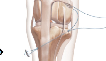
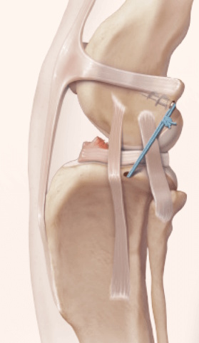
Extra-Capsular Stabilization - additional information
Post Surgical Radiographs
While the patient is still anesthetized, the patient is taken into radiology for post-surgery radiographs (X-rays). The radiographs are assessed to ensure the apparatus FASTak screw and toggle are appropriately positioned.
Recovery From Anesthesia
After the post-surgical radiographs have been reviewed by the surgeon, the patient is moved to recovery. The patient's leg is cleaned, and a light, padded bandage is applied to the limb to help prevent post-surgical swelling.
The patient is extubated and moved to a recovery kennel. The epidural (bupivacaine and morphine) provides 6 hours of post-surgical relief for the patient. This equates to 3-4 hours of pain relief after the patient has woken up from anesthesia. After the epidural has worn off, the patient will be able to walk out to the bathroom and already be able to place weight on their operated limb. Most patients will feel good enough to enjoy a nice dinner after surgery.
Post Surgical Care
For routine procedures, the usual hospital stay is 2-3 days. We often board patients for a longer duration after surgery when their owners feel they are unable to provide the necessary care required during the recovery period. A light, padded bandage is applied to the operated limb to help reduce post-surgical bruising and swelling.
Typical rechecks are at 2, 6, and 12 weeks.
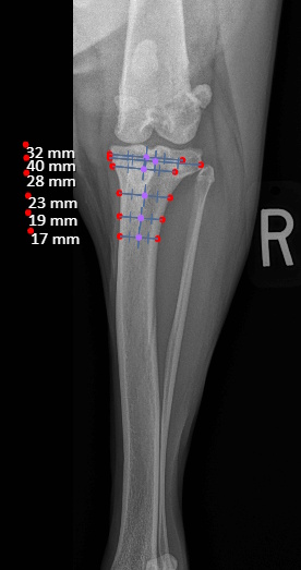
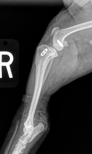
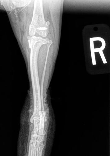
Tibial Plateau Leveling Osteotomy (TPLO) - additional information
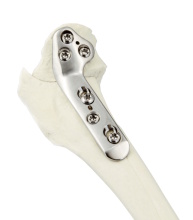
An osteotomy is a surgical cut in a bone. In the cruciate-deficient stifle, a cut is made in the top of the tibia. The top portion of the bone that contains the articular surface is rotated to level it. The two pieces of bone are held in place with a metal plate and screws.
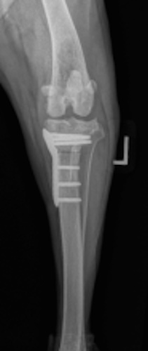
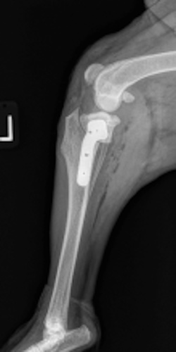
Post Surgical Radiographs
While the patient is still anesthetized, the patient is taken into radiology for post-surgery radiographs (X-rays). The radiographs are assessed to measure the new tibial plateau angle. We are aiming for 5-6 degrees relative to the long axis of the tibia. The apparatus (plate and screws) are assessed for size and appropriate position.
Recovery From Anesthesia
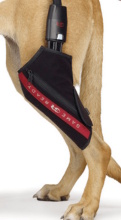 After the post-surgical radiographs have been reviewed by the surgeon, the patient is moved through to recovery. The patient's leg is cleaned, and a sports wrap that provides cold compression therapy (Game Ready) is applied to the operated limb for 30 minutes.
The patient is extubated and moved through to a recovery kennel. The epidural (bupivacaine and morphine) provides 6 hours of post-surgical relief for the patient. This equates to 3-4 hours of pain relief after the patient has woken up from anesthesia. After the epidural has worn off, the patient will be able to walk out to the bathroom and already be able to place weight on their operated limb. Most patients will feel good enough to enjoy a nice dinner after surgery.
Post Surgical Care
For routine procedures, the usual hospital stay is 2-3 days. We often board patients for a longer duration after surgery when their owners feel they are unable to provide the necessary care required during the recovery period.
Typical rechecks are at 2, 6 and 12 weeks. Patients come back at two weeks to have their sutures removed. Until the sutures are removed the patients will wear an E-Collar (cone of shame) to prevent them from licking at their incision. The inflatable collars do not provide sufficient protection for surgery on the knee and are not recommended.
For the first 6 weeks after surgery, the patient will need to be confined to a small kennel or crate to prevent them from over-exercising. For the first two weeks after surgery, the patients will need to be taken out for toilet breaks (on a leash) and then returned to their crate. At two weeks, the patient will have their sutures removed, and they no longer need to wear the E-collar.
At two weeks after surgery the pets will be able to start going on short walks whilst being confined to their leash.
At six weeks after surgery, we will take radiographs to confirm the healing process is progressing as expected. The patient will then be able to move around within the confines of the house without being kept in a crate. Outside, they will still need to be kept on a leash for an additional 6 weeks. At 12 weeks after surgery, a final recheck exam and series of radiographs will be obtained. The patients will be cleared at this visit to resume activity off-leash.
Additional Types of Orthopedic Injuries and Surgeries
Carpal Hyperextension
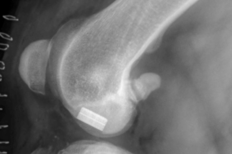
Stifle (knee) Osteochondritis Dissecans (OCD)
Osteochondritis dissecans (OCD) is a developmental condition that arises due to a disturbance in the normal differentiation of cartilage cells resulting in failure of endochondral ossification (essential process during foetal development of skeletal system resulting in bone formation).
In dogs that grow very quickly, the rapid cartilage growth can outstrip its own blood supply causing abnormal cartilage development resulting in lameness, pain and subsequent osteoarthritis. In some cases, flaps of diseased cartilage become separated from the remaining cartilage surface. This is called osteochondritis dissecans.
How Do I Know If MY Dog Has OCD?
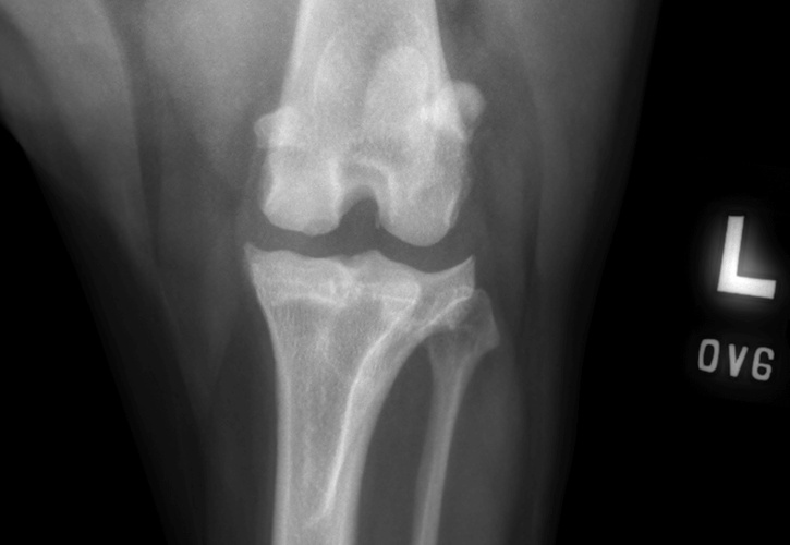
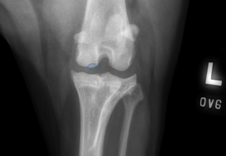
OCD lesion of the canine stifle highlighted with a blue dotted line
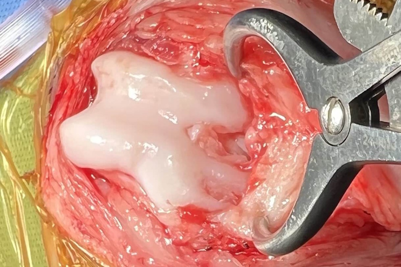
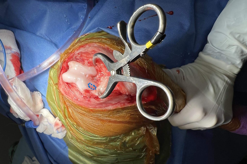
How Is Stifle OCD Treated?
- Surgical Removal of the Cartilage Flap
- SynaCart
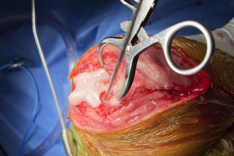
SynaCart
Manufactured by Arthrex, SynaCart™ represents the cutting edge of treatment for canine OCD. The implant is a second generation synthetic osteochondral resurfacing implant which incorporates the polycarbonate urethane of the SOR implant mentioned previously and combines this with a trabecular titanium metal which allows bone to grow into the implant. Synacart was developed by Arthrex in conjunction with Dr Noel Fitzpatrick who was one of Dr Ned Trathans mentors back in the UK.

Cruciate Ligament Disease (CCL)
Patellar Luxation
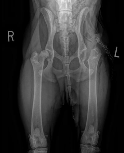
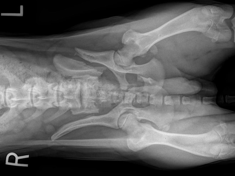
Hip Dysplasia
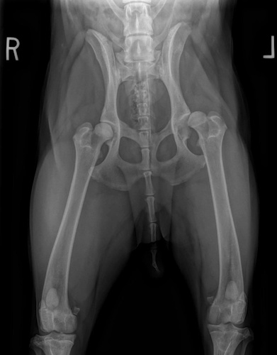
Hip Dysplasia in canines
Surgical Treatments of Hip Dysplasia
- Juvenile Pubic Symphysiodesis (JPS)
- Triple Pelvic Osteotomy (TPO)
- Total Hip Replacement (THR)
- Femoral Head and Neck Excision (FHNE)
Coxofemoral Luxation (Hip Dislocation)
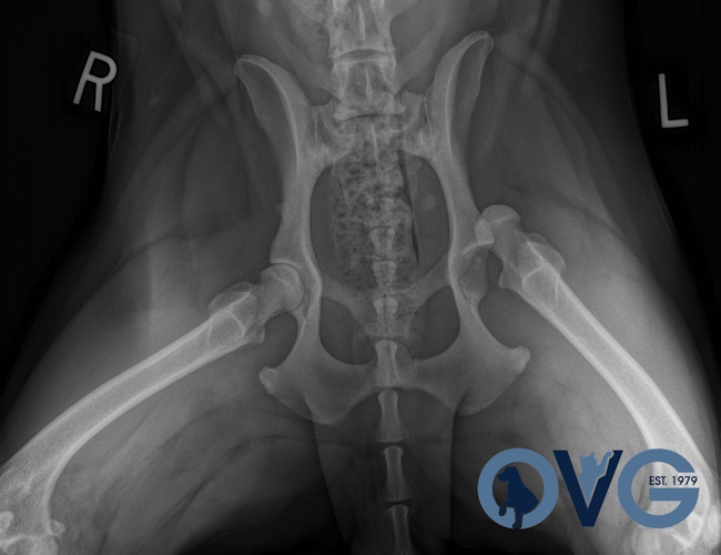
Hip Dislocation in canines
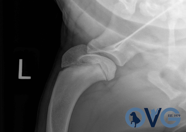
Treatment Option for Hip Dislocation
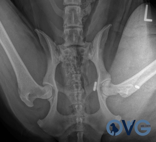
Post-op of a Hip Replacement
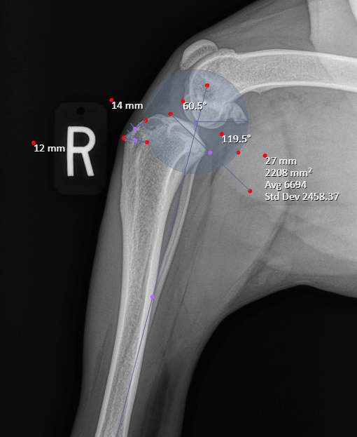
Shoulder Osteochondritis Dissecans (OCD)
Shoulder Osteochondritis Dissecans (OCD)
How Is Shoulder OCD Treated?
- Surgical Removal of the Cartilage Flap
- SynaCart
SynaCart
Manufactured by Arthrex, SynaCart™ represents the cutting edge of treatment for canine OCD. The implant is a second generation synthetic osteochondral resurfacing implant which incorporates the polycarbonate urethane of the SOR implant mentioned previously and combines this with a trabecular titanium metal which allows bone to grow into the implant. Synacart was developed by Arthrex in conjunction with Dr Noel Fitzpatrick who was one of Dr Ned Trathans mentors back in the UK.
Shoulder Osteochondritis Dissecans (OCD) with a SynaCart Implant
Types of Fractures
Fore Limb (Humerus, Radius/Ulnar, Metacarpus)
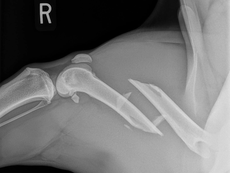
Humerus
Humerus
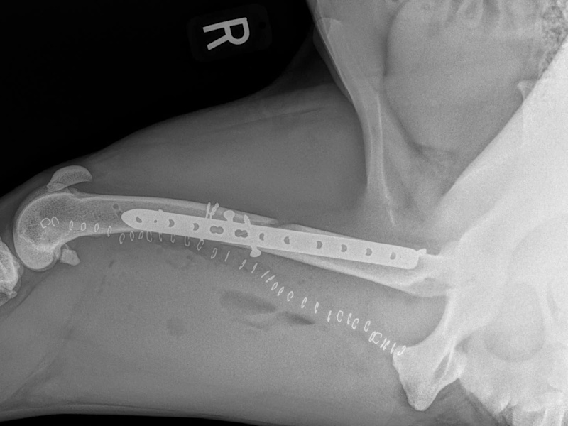
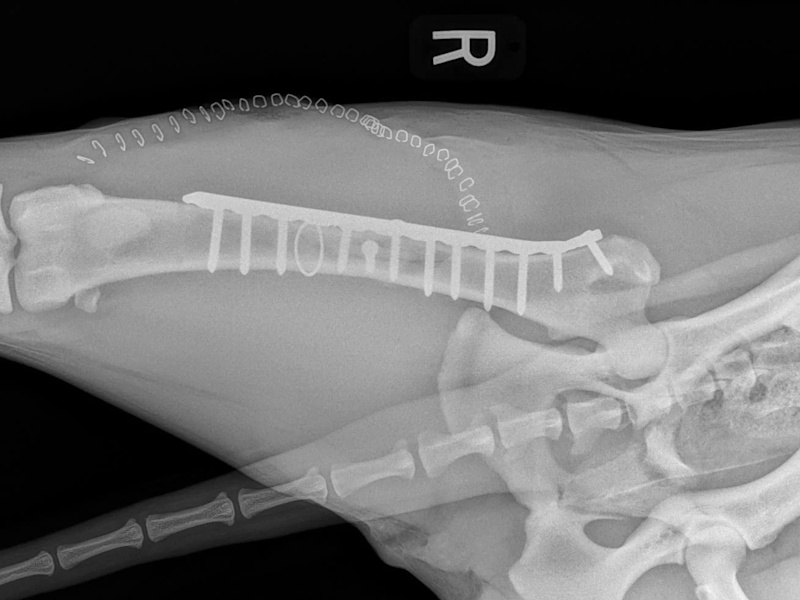
Repaired
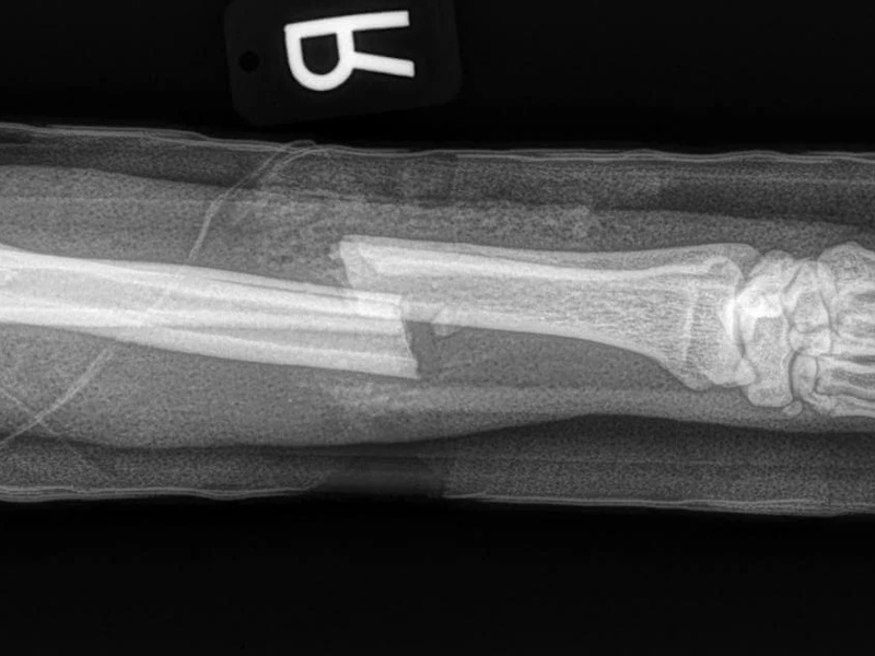
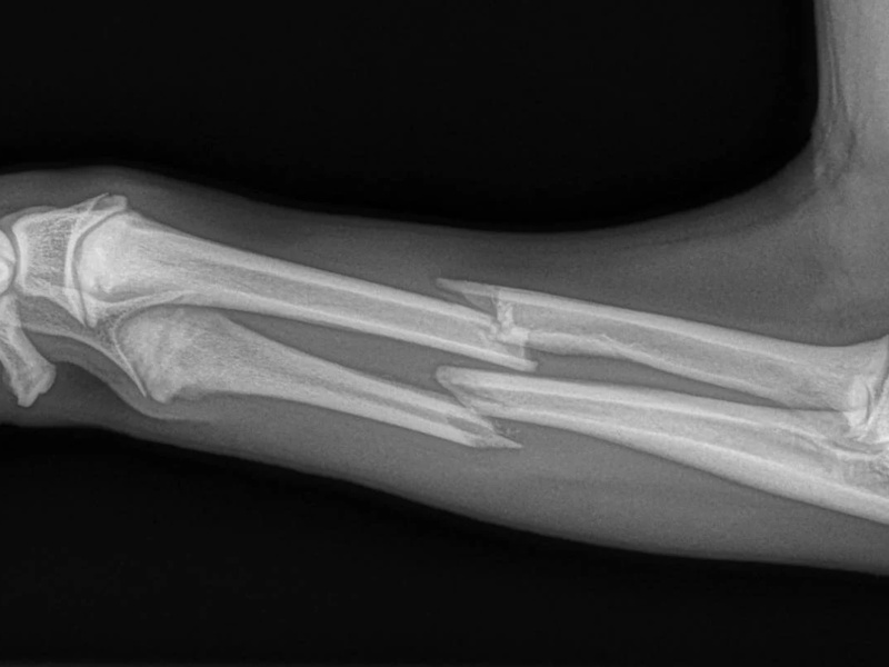
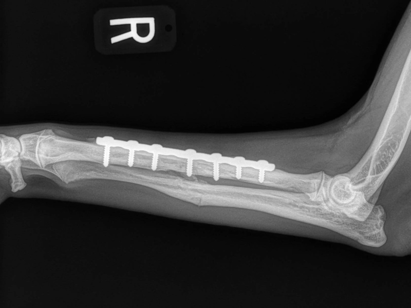
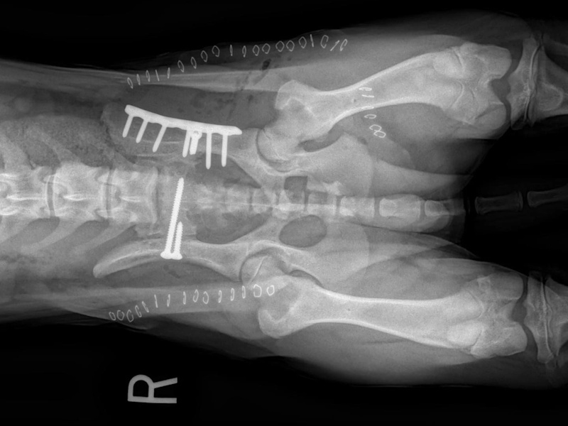
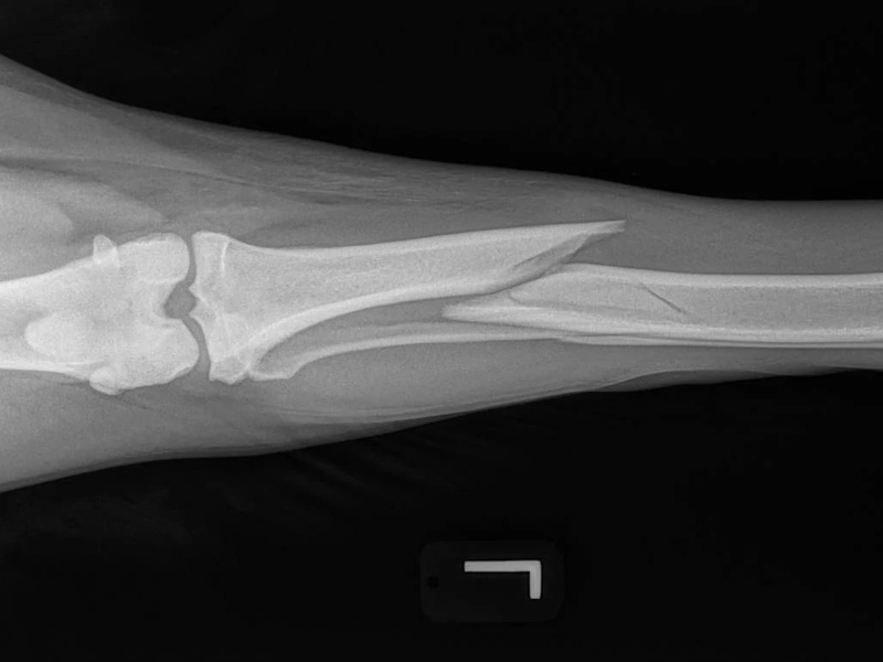
Radius/Ulnar
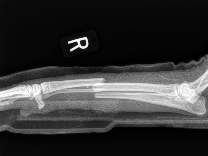
Radius/Ulnar
Radius/Ulnar
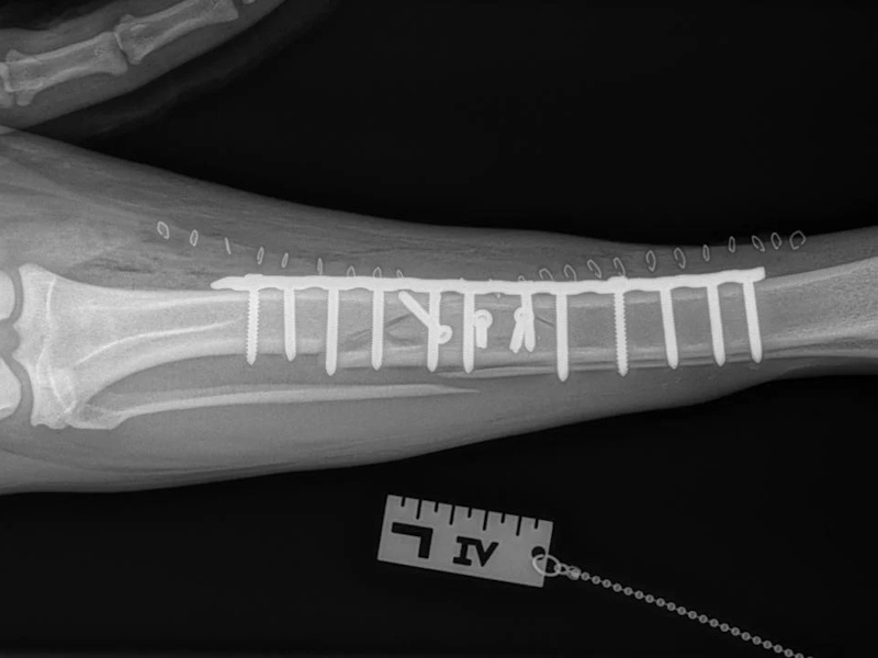
Plated
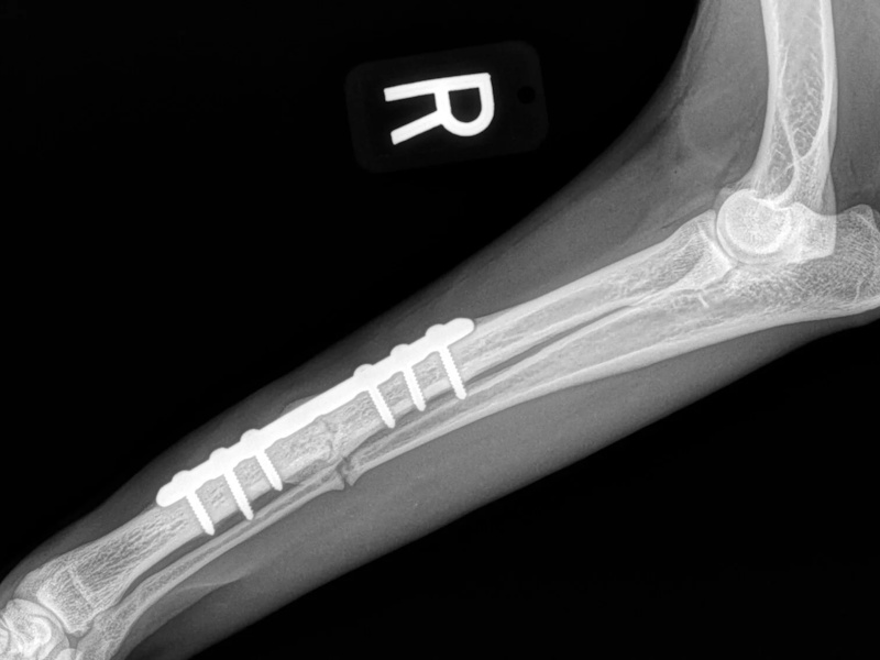
Radius/Ulnar
Radius/Ulnar
Radial plate and Ulnar pin
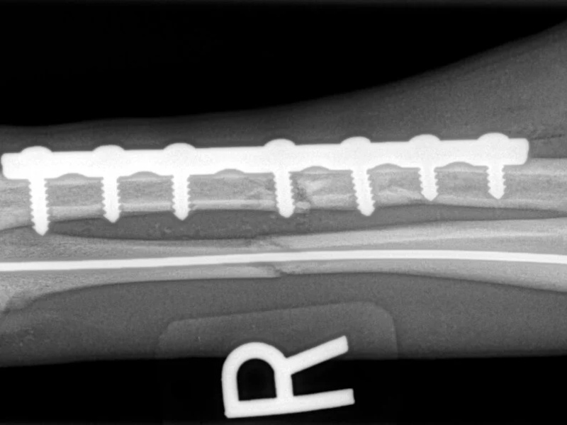
Radius/Ulnar
Radial plate with Ulnar pin removed
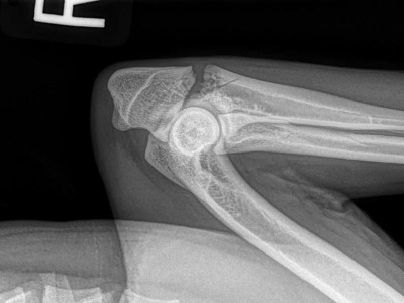
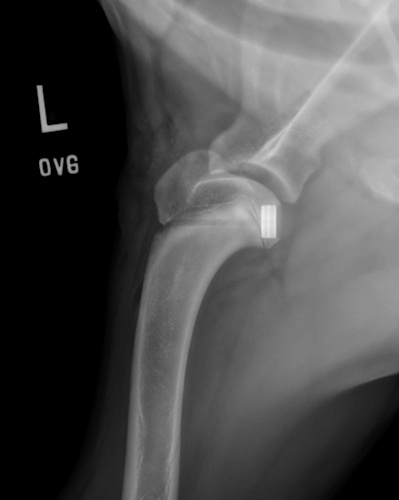
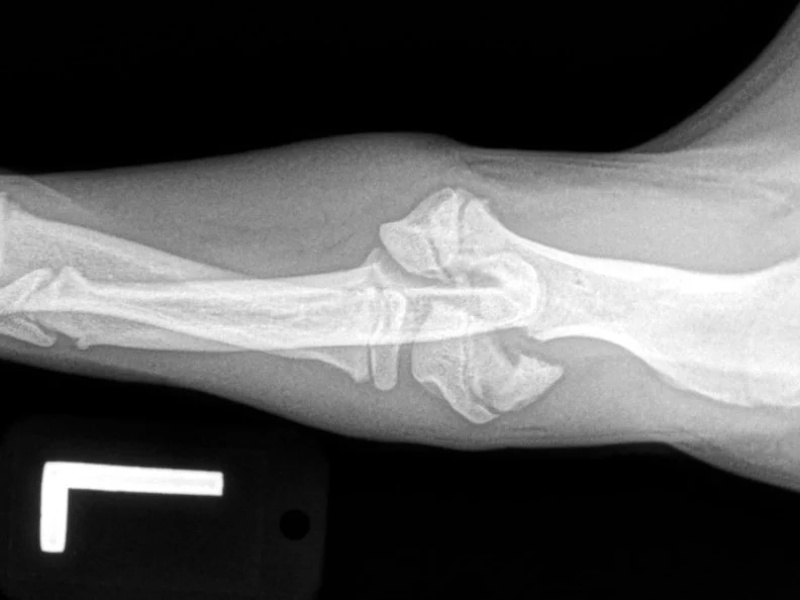
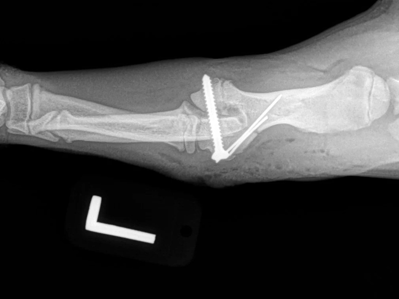
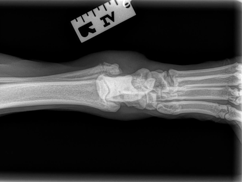
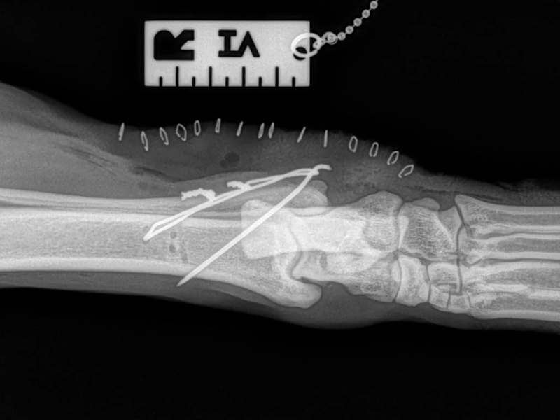
Ulnar articular
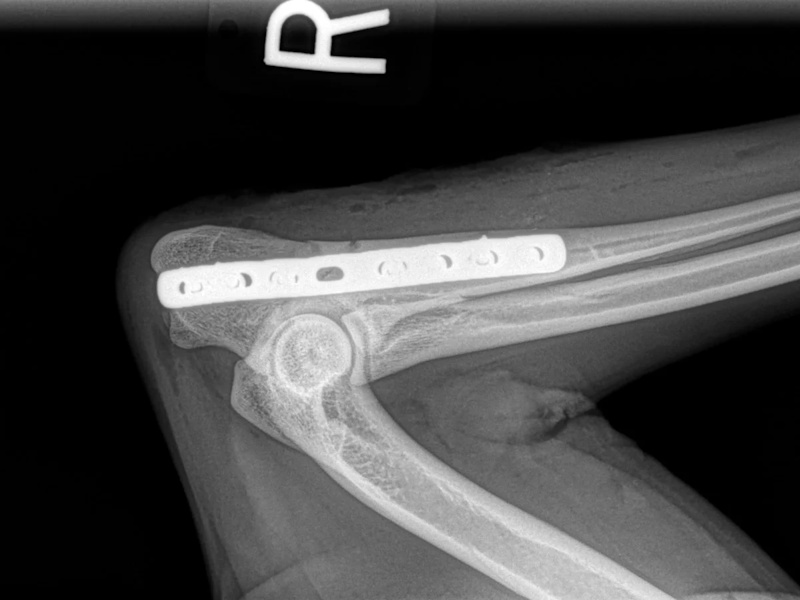
Ulnar articular
Plated
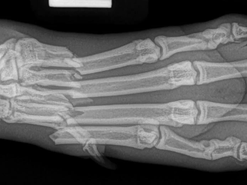
Metacarpal
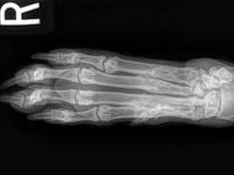
Metacarpal
Repaired
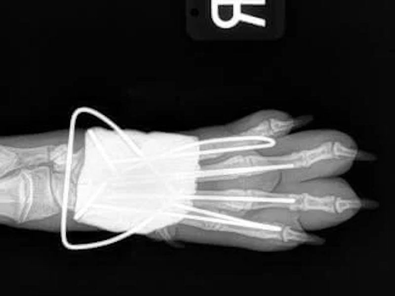
Metacarpal
Spider Fixator
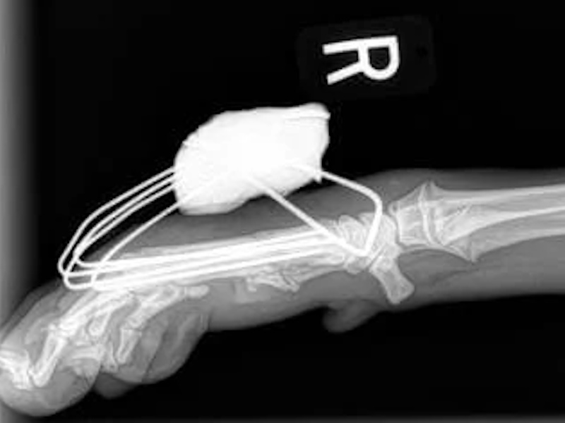
Metacarpal
Spider Fixator
Hind Limb (Pelvis, Femur, Patella, Tibia/Fibula, Lateral Malleolus)
Pelvis
Pelvis
Plate repaired
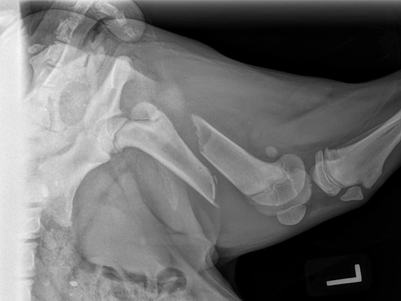
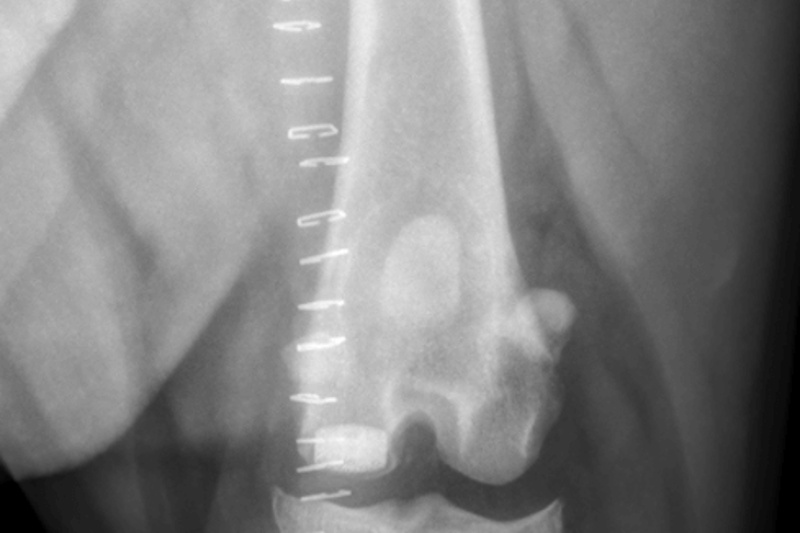
Femur
Femur
Plate repaired
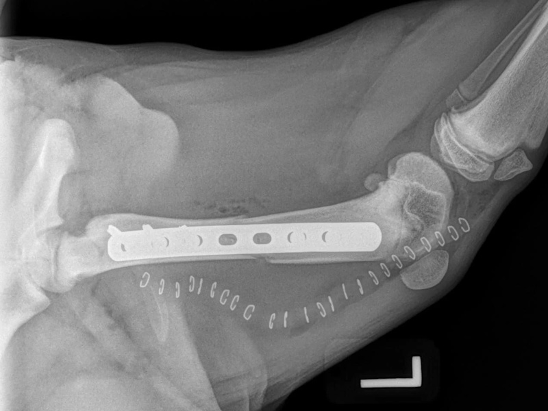
Femur
Plate repaired
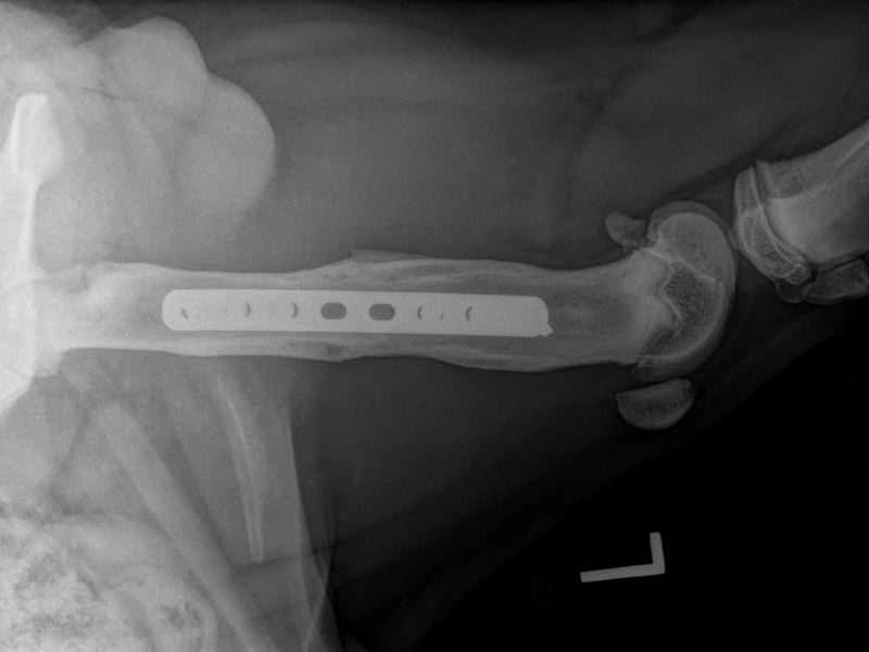
Femur
Femur
Plate repaired
Femur
Plate repaired
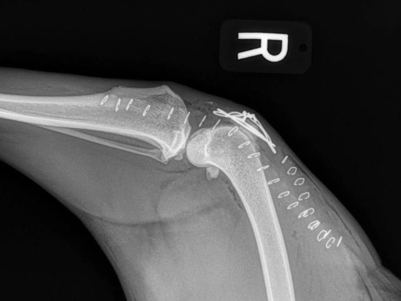
Patella
Repaired
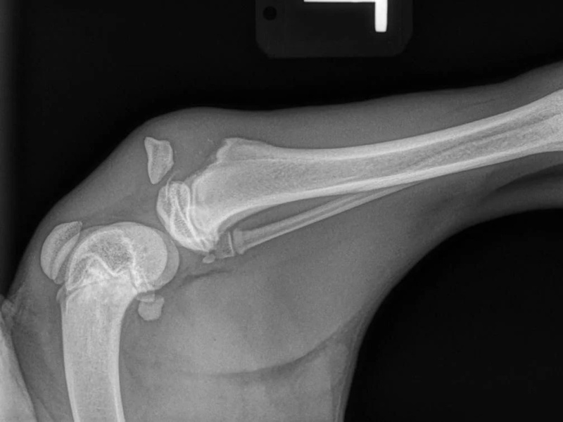
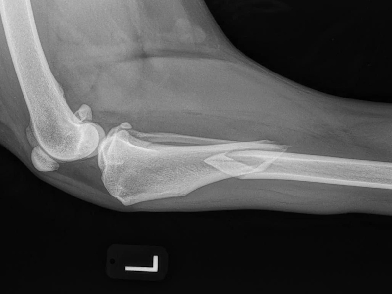
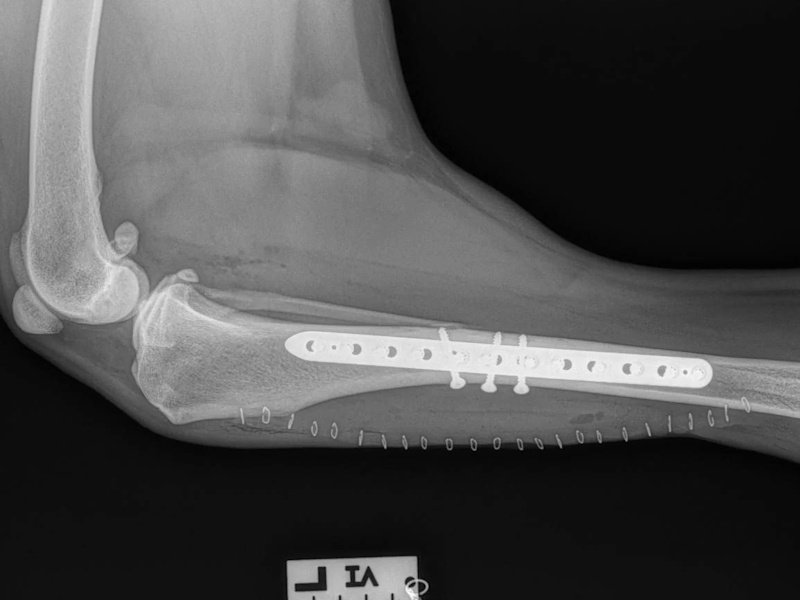
Tibia
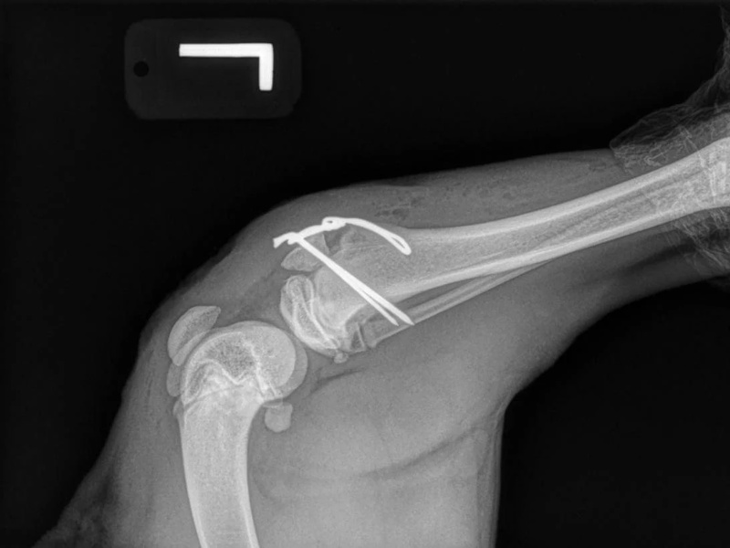
Tibia
Repaired with pin and tension band
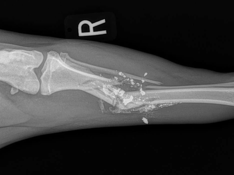
Tibia
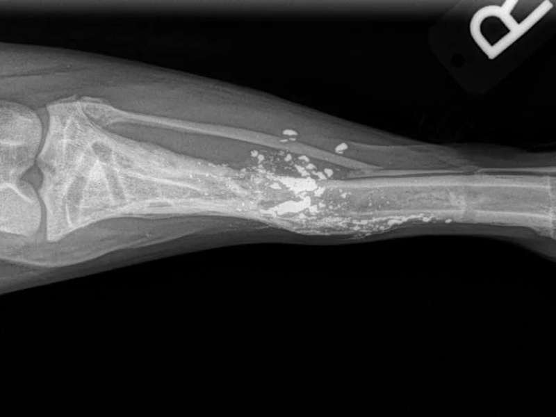
Gun shot wound
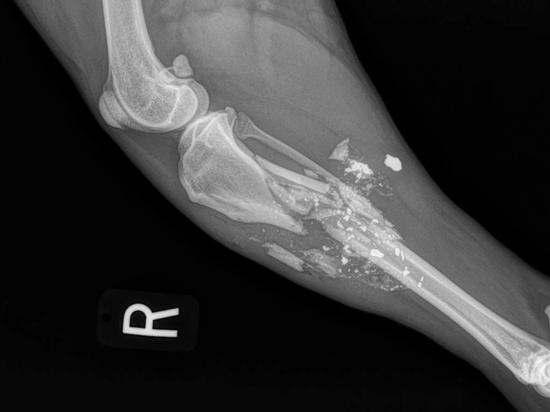
Tibia
Gun shot wound
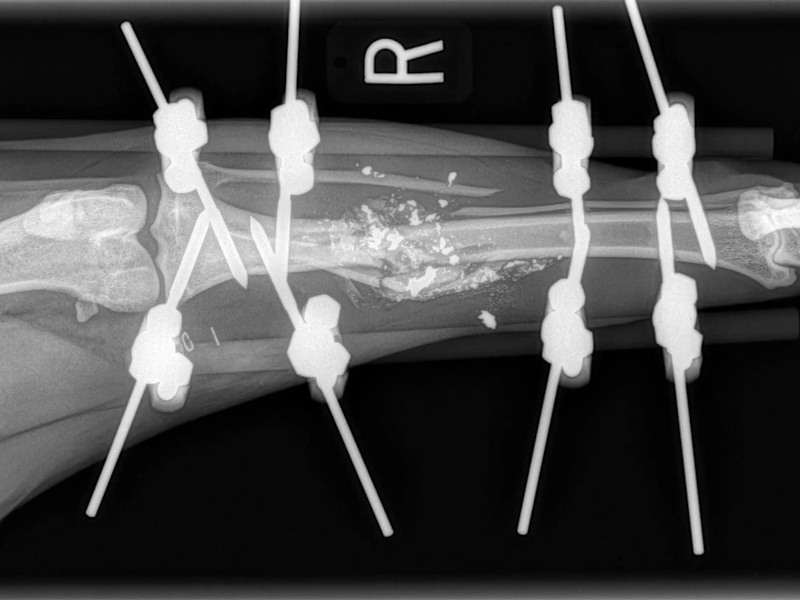
Tibia
External fixator
Tibia
Repaired
Tibia
Tibia
Plate repaired
Tibia
Tibia
Plate repaired
Lateral Malleolus
Lateral Malleolus
Repair pin and tension band